6.
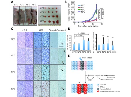
HS enhanced tumorigenesis in a xenograft model. (A) Flank tumors were established in BALB/c nu/nu female mice (n = 4 per group) as described in materials and methods. Mice were killed at day 12 after flank injection. Representative tumors were showed. The 45°C and 48°C preheated CT-26 cells resulted in a dramatic rise of tumor weight. (B) Growth curves of xenograft tumors after the injection of pre-treated and untreated cells in mice (P< 0.01). (C) Representative hematoxylin and eosin (HE)-staining and IHC staining (Ki67, clevead-caspase3) images of xenograft tumors harvested from mice that received subcutaneous injection of treated and untreated CT-26 cells. (Magnification: left half is 100 ×, right half part is 200 ×) (D) The numbers of Ki67+ and cleved-caspase-3+ cells were counted and analyzed with unpaired Student’st-test. (E) Schematic graphs showed HS induced proliferation, invasion and stemness pluripotency related cascade. All data were from three independent experiments.

