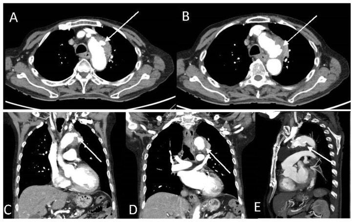
Figure 3.
An 89-year-old male with peri-aortic pseudoaneurysms.
Findings: Initial CT aortogram showed that the periaortic soft tissue mass along the lateral wall of the aortic isthmus appeared less prominent. However, there were two new contrast-filled focal out-pouching's along the lateral and inferior wall of the aortic isthmus. Axial images of the two small pseudoaneurysms along the lateral (A, arrow) and inferior (B, arrow) wall of the aortic arch measuring 0.7 x 1.1 cm and 1.7 x 1.3 cm, respectively. Coronal images of the two small pseudoaneurysms along the lateral (C, arrow) and inferior (D, arrow) wall of the aortic arch. (D) Sagittal image shows the inferior pseudoaneurysm (arrow).
Technique: Contrast enhanced CT aortogram with axial, coronal and sagittal reformats. CT scan settings were 3.00 mm slice thickness at 100 kV and 115 mAs.

