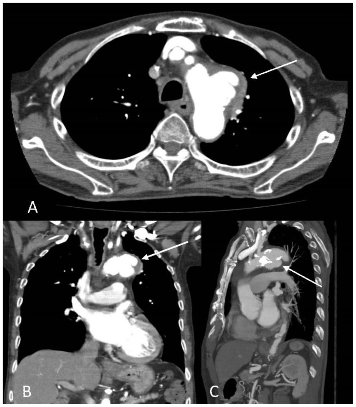
Figure 4.
An 89-year-old male with enlarging peri-aortic pseudoaneurysms.
Findings: The follow-up CT aortogram showed the previously described small pseudoaneurysms along the lateral and inferior wall had coalesced and developed into a larger lobulated pseudoaneurysm with a wide neck measuring 4.0 cm and pseudoaneurysm sac measuring 3.9 x 2.3 x 3.5 cm (A, Axial images, B, coronal images and (C) sagittal image arrows). In view of increasing size of the pseudoaneurysm the patient underwent urgent TEVAR.
Technique: Contrast enhanced CT aortogram with axial, coronal and sagittal reformats. CT scan settings were 3.00 mm slice thickness at 120 kV and 70 mAs.

