
Figure 2:
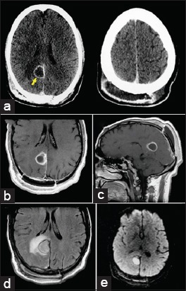
Postoperative axial computed tomography following contrast administration. (a) Revealing a subgaleal extracranial homogenous low-density lesion with rim enhancement (right). There was also another intracranial ring-enhancing collection at the site of the tumor resection cavity (left), suggesting a brain abscess (arrow). Postoperative magnetic resonance imaging showing both extracranial and parafalcine cystic masses with homogenous, low-intensity signal on T1-weighted images. (b and c) And high-intensity signal on the fluid-attenuated inversion recovery sequence. (d) There was peripheral enhancement after gadolinium injection (b and c) and significant cerebral edema surrounding the resection cavity. (d) Note the restricted diffusion-weighted image (bright signal) of the extracranial and parafalcine collections (e).