
Figure 1:
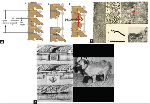
(a) Proposed model of removal of the injured segment (star), transection of the cord above and below (ovoids) and fusion with polyethylene glycol (arrow) along with vertebral shortening and stabilization (adapted from Qiu et al., 2015). (b) Box 1: Freeman discovered that regrowing fibers could be made to grow across the sectional interface in rats, dogs and monkeys and that this translated into electrophysiological transmission and behavioral recovery. (c) Box 1: Since spinal cord transection is not common, Freeman reasoned that he could leverage his technique of spinal cord regeneration in clinical models of spinal cord injury by cleanly cutting the cord above and below the level of injury, removing the injured segment of the cord (2 cm), doing a vertebrectomy and bringing the two fresh ends of the cord together and holding them in place with plasma clot before the dura mater was tightly closed. Dogs could thus be made to rewalk for short distances (the one displayed had almost two thoracic segments removed).