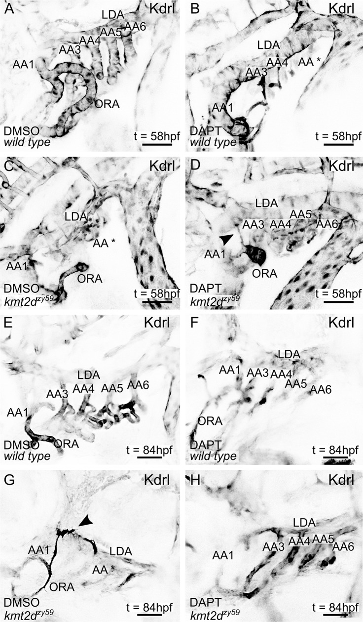
Fig 9. Pharmacological inhibition of Notch signaling rescues early endothelial phenotypes and suppresses ectopic blood vessel formation in zebrafish kmt2d mutants.
(A–H) Still images (MIP) from time-lapse live imaging performed from 2 dpf to 2.5 dpf (A–D) and 3 dpf to 3.5 dpf (E–H). wild-type tg(Kdrl:GFP) and kmt2dzy59;tg(Kdrl:GFP) samples were treated with DMSO (n = 12; A, C, E, and G) or DAPT (n = 12; B, D, F, and H) from 1 dpf to 2 dpf. After treatment, samples were washed and prepared for in vivo time-lapse imaging. Cranial-lateral view at the level of AA development corresponding to video as follow: A, S7 Video; B, S9 Video; C, S8 Video; D, S10 Video; E, S11 Video; F, S13 Video; G, S12 Video; H, S14 Video. Images were selected at last time points recorded. Videos and images were converted to grayscale and inverted for better visualization. Asterisk (B, C, and G = AA*) denotes abnormal or missing vascular development of AA sprouts at the level of the ventral border of LDA. (scale bars = 50 μm). AA, aortic arch; AA1, mandibular arch; AA2, hyoid arch; AA3, first branchial arch; AA4, second branchial arch; AA5 third branchial arch; AA6, fourth branchial arch; dpf, days post fertilization; hpf, hours post fertilization; kdrl, kinase insert domain receptor like; LDA, lateral ventral aorta; MIP, maximum intensity projection; ORA, opercular artery.

