Figure 5.
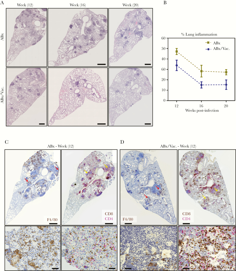
Adjunct respiratory mucosal immunotherapy reduces tuberculosis-associated tissue pathology and enhances CD8 T cell infiltration in the lung during antibiotic continuation phase. Experiments were set up as depicted in experimental schema Figure 3A. (A) Representative micrographs of lung sections stained with hematoxylin and eosin, comparing the extent of lung inflammation and granulomatous lesions at weeks 12, 16, and 20 weeks postinfection. Scale bars indicate 500 µm. (B) Line graph showing semiquantified area of lung inflammation. Displayed values are averages from 3 micrographs per mouse. (C and D) Representative micrographs of immunohistochemically stained lung sections at 12 weeks postinfection visualizing the spatial distribution of F4/80 macrophages (brown stain) and of CD4 (red stain) and CD8 (brown stain) T cells costained in consecutive sections. Red arrows highlight macrophage-rich areas. Yellow arrows highlight T cell-rich areas. Top panel scale bars indicate 500 µm; bottom panel scale bars indicate 50 µm. ABx, antibiotics alone.

