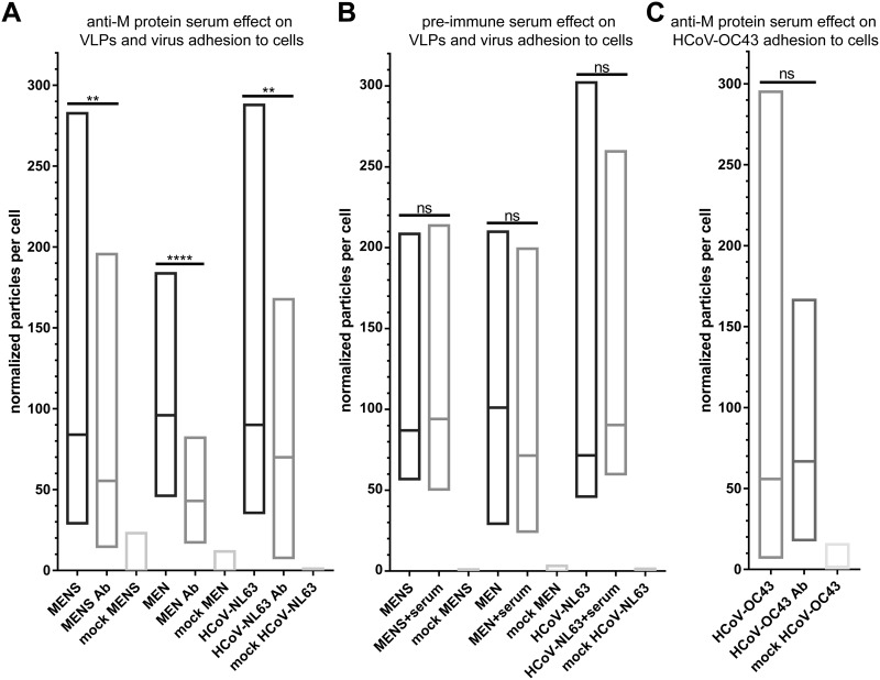
FIG 8.
VLP and HCoV-NL63 pseudoneutralization assay. (A and B) LLC-Mk2 cells were inoculated with MEN or MENS VLPs or HCoV-NL63 preincubated with anti-M serum, denoted “Ab” (A), or the respective preimmune serum (B). (C) The specificity of anti-M serum was examined by testing its effect on HCoV-OC43 adhesion to HRT-18G cells. Cells fixed and stained with anti-N monoclonal antibody were visualized using confocal microscopy. The number of particles and number of cells were quantified using the ImageJ Fiji 3D Objects Counter tool. The number of particles per cell is presented as a minimum-maximum graph with a line set at the median value. Each bar shows data from a minimum of 24 212- by 212-μm fields of view, registered from three different samples (**, P < 0.01; ****, P < 0.0001).

