FIG 2.
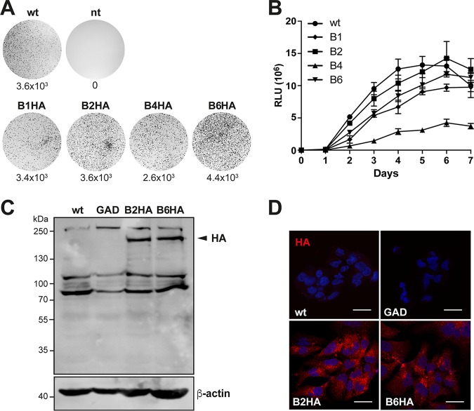
Replication of HA-tagged subgenomic HEV replicons. (A) Replication efficiency of HA-tagged subgenomic HEV replicons. Hep293TT cells were fixed 12 days postelectroporation with in vitro-transcribed RNA from a subgenomic replicon construct (pUC-HEV83-2_Neo_B1-HA, pUC-HEV83-2_Neo_B2-HA, pUC-HEV83-2_Neo_B4-HA, or pUC-HEV83-2_Neo_B6-HA) and stained with crystal violet. The parental HEV83-2-27_Neo replicon (wild type [wt]) and nontransfected cells (nt) served as positive and negative controls, respectively. Results from a representative experiment are shown, with the number of colonies per microgram of transfected RNA indicated below each plate. (B) Culture supernatants from Hep293TT cells electroporated with in vitro-transcribed RNA from the parental pUC-HEV83-2_Gluc (wild type [wt]) or the pUC-HEV83-2_Gluc-derived replicon construct harboring 15-nucleotide transposon insertions in site B1, B2, B4, or B6 were analyzed for luciferase activity over 7 days. Gaussia luciferase activity was measured by adding 10 μl of supernatant from Hep293TT cells electroporated with luciferase HEV replicon constructs to 60 μl of substrate buffer (0.8 μM coelenterazine–PBS). Relative light units (RLU) were determined four times for each time point. (C and D) Immunoblotting (C) and immunofluorescence detection (D) of HA-tagged ORF1 protein in Hep293TT cells. Cells were electroporated with wild-type (wt), polymerase-defective (GAD), or HA-tagged (B2HA and B6HA) selectable subgenomic replicon constructs. (C) Protein lysates were prepared 5 days postelectroporation and separated by 8% SDS-PAGE, followed by sequential immunoblotting using rabbit MAb C29F4 against HA and mouse MAb AC-15 against β-actin. The arrowhead denotes HA-tagged ORF1 protein. Molecular weight markers are indicated on the left. (D) Cells were fixed 5 days postelectroporation and subjected to immunofluorescence with rabbit MAb C29F4 against HA as primary antibody and Alexa Fluor 594 anti-rabbit IgG as secondary antibody. Cell nuclei were counterstained with DAPI. The scale bar represents 20 μm.

