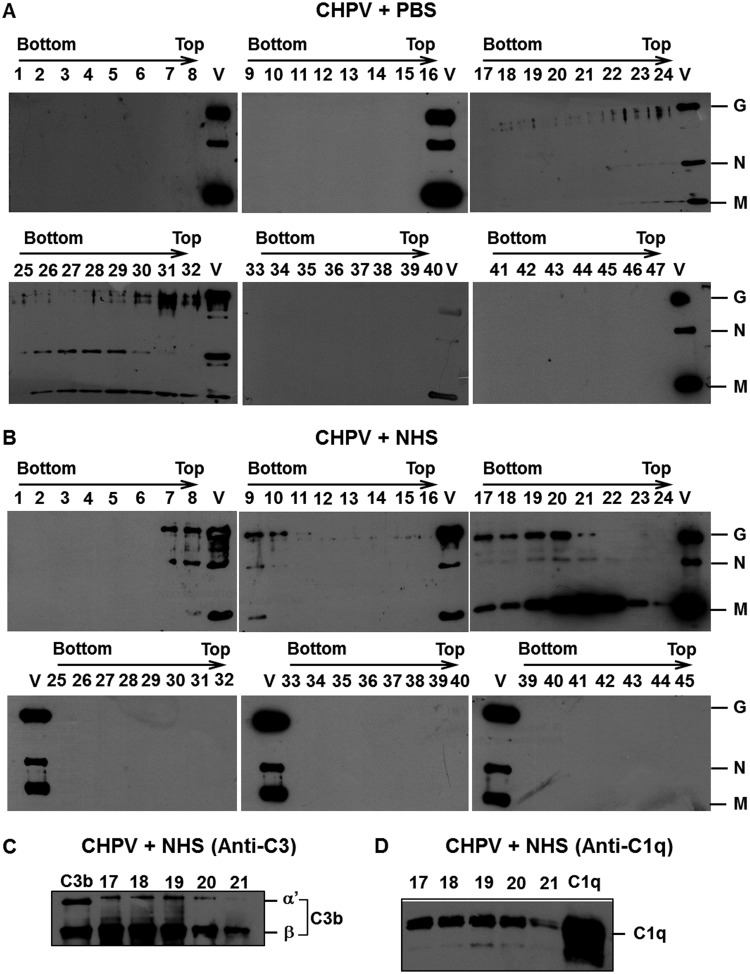
FIG 7.
Differential shift in Chandipura virus fractions upon NHS treatment is due to association of complement components. Gradient-purified CHPV was incubated with PBS or NHS (1:5) and layered on top of a 15% to 60% linear sucrose gradient and subjected to ultracentrifugation. Western blot analysis was carried out on the fractions collected. Panel A represents the CHPV+PBS samples (fraction 25 to 32 shows reactivity to anti-CHPV antibodies), while blots in panel B represent the CHPV+NHS samples (fractions 17 to 24 are reactive to anti-CHPV antibodies). The letter V in every blot represents purified CHPV run as a control. The labels G, N, and M indicate CHPV proteins glycoprotein G, nucleocapsid, and matrix protein, respectively. The peak fractions were also probed to detect complement component C3 (C) and C1q (D) association. Purified proteins C3b and C1q were used as controls and are labeled thus in the blots.

