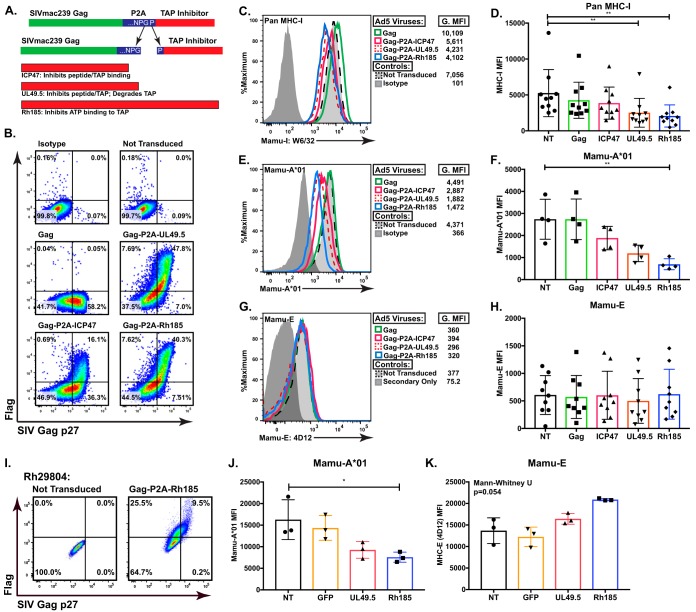
FIG 1.
ICP47, UL49.5, and Rh185 inhibit TAP function in rhesus macaque Cells. (A) Schematic of three vector inserts inclusive of SIVmac239 Gag and the Flag-tagged TAP inhibitors ICP47 (HSV-2), UL49.5 (BHV-1), or Rh185 (RhCMV), separated by 2A peptide derived by porcine teschovirus-1. The resulting cleavage during peptide synthesis yields an additional 18 amino acids attached at the C terminus of Gag but only one proline added to the N terminus of the selected TAP inhibitor. (B) Representative flow cytometry data of SIVmac239 Gag p27 and Flag-tagged TAP inhibitor expression in BLCL transduced at a multiplicity of infection of 2,000 with Ad5/Gag, Gag-P2A-ICP47, Gag-P2A-UL49.5, or Gag-P2A-Rh185. (C and D) Staining of BLCL with pan-MHC-I antibody (W6/32) showed that MHC-I expression was significantly suppressed as a consequence of TAP inhibition. G.MFI, geometric mean fluorescence intensity. Panel C shows representative flow cytometry data from one animal, while panel D shows data compiled from multiple RM. (**, P < 0.001). (E and F) Subset analysis on A*01 surface expression using RM that were positive for the MHC-Ia allele Mamu-A*01. After transduction with Ad5 viral vectors, this subset was stained with an antibody specific to Mamu-A*01. With the smaller subset, only Rh185 transduced BLCL reach statistical significance (**, P < 0.001). Panel E shows representative flow cytometry data from one animal, while panel F shows data compiled from multiple RM. (**, P < 0.001). (G and H) The BLCL from the experiments described in panels C and D were stained with the MHC-E-specific 4D12 antibody, and surface MHC-E was assessed. Panel G shows representative flow cytometry data from one animal, while panel H shows compiled data from multiple RM. (I) Representative flow cytometry data of SIVmac239 Gag p27 and Flag-tagged TAP inhibitor expression in monocyte-derived macrophages (MDM) transduced at a multiplicity of infection of 2,000 with Ad5/Gag-P2A-Rh185, Ad5/Gag-P2A-UL49.5, or Ad5 expressing green fluorescent protein (GFP). Posttransduction MDM from Mamu-A*01-positive RM were stained for surface Mamu-A*01 expression (J) and surface Mamu-E expression (K). Values shown in panels D, F, H, J, and K are background subtracted. NT, not transduced.

