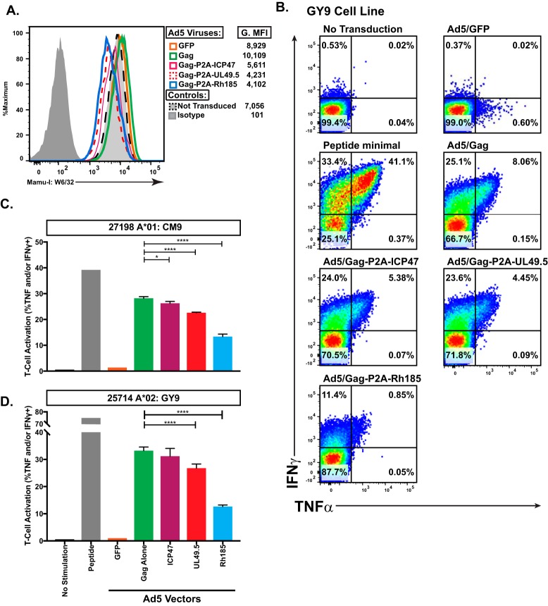
FIG 2.
MHC-Ia-restricted T cells have reduced recognition of TAP-inhibited targets. (A) Surface MHC-I levels on the Ad5 transduced heterologous BLCL (Mamu-A*01/A*02+) used as effectors to test the impact of TAP inhibition on MHC-Ia-restricted CD8+ T cell recognition. G.MFI, geometric mean fluorescence intensity. (B) Representative ICS data of Gag GY9-specific CD8+ T cells in response to coincubation with the antigen-presenting cells indicated above each plot. (C) Activation of Gag CM9-specific CD8+ T cells as measured by secretion of TNF-α and/or IFN-γ in response to coincubation with exogenous peptide or BLCL antigen-presenting cells infected with the indicated Ad5 vectors (*, P < 0.01; ****, P < 0.0001). (D) Activation of Gag GY9-specific CD8+ T cells as measured by secretion of TNF-α and/or IFN-γ, in response to coincubation with exogenous peptide or BLCL antigen-presenting cells infected with the indicated Ad5 vectors (*, P < 0.01; ****, P < 0.0001).

