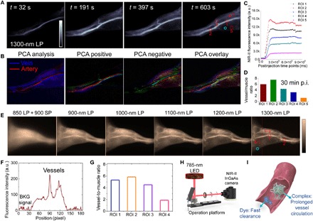
Fig. 3. The IR-783@BSA complex enables high-contrast, real-time NIR-II vessel imaging.

(A) The recorded NIR-II video of hindlimb vessels in mouse at several time points p.i. of the IR-783@BSA complex at 1300-nm long-pass (LP) filters. (B) PCA analysis from the real-time video imaging distinguished both vein and artery vessels, as well as small vessels and background. (C) The monitor of fluorescence intensity profile of five selected vessels in NIR-II video imaging of (A). High contrast between the vessel and background is maintained for up to ~30 min in the NIR-II imaging window. The red dotted square is the ROI of vessel, and the cyan circle is the ROI of muscle. a.u., arbitrary units. (D) Vessel-to-muscle signal ratio at 30 min p.i. showed high-contrast value up to 6. (E) The high brightness of the IR-783@BSA complex affords low-power LED-excited hindlimb imaging at wide NIR-II subwindows. The low-power LED light (10 to 50 mW/cm2) is more suitable for potential clinical use. The dotted line shows a representative cross-sectional profile of hindlimb. SP, short-pass. (F) The cross-sectional intensity profile of hindlimb in (E) (the dotted line). BKG, background. (G) LED-excited imaging shows equal vessel-to-muscle signal ratio [ROI 1 to 4 in (E)] with 785-nm laser excited imaging. (H) Scheme of LED-excited NIR-II imaging system. (I) Scheme of prolonged vessel circulation of the IR-783@BSA complex compared with free IR-783.
