
Fig. 4. NIR-II microscope imaging of the IR-783@BSA complex affords high-performance vessel imaging with both high resolution and contrast.
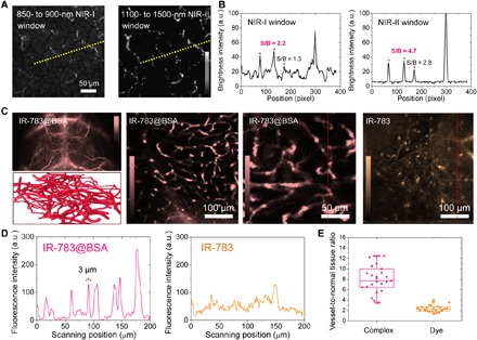
(A) Ex vivo NIR-II microscope imaging of mouse brains at 1 hour p.i. of IR-783@BSA at both NIR-I and NIR-II windows. (B) Cross-sectional intensity profile of both NIR-I and NIR-II images at the same position. NIR-II imaging affords two times enhancement of SBR. (C) In vivo NIR-II whole-body (2.5×), vessel scheme and ex vivo microscope imaging of shaved/sectioned mouse head/brains after IR-783@BSA complex administration over 1300- and 1200-nm long-pass filters, respectively. (D) Cross-sectional intensity profile of both IR-783@BSA complex and free IR-783 images. (E) Vessel–to–normal tissue ratio statistics of both IR-783@BSA complex and free IR-783 images indicated that the IR-783@BSA complex exhibited super vessel imaging capacity.