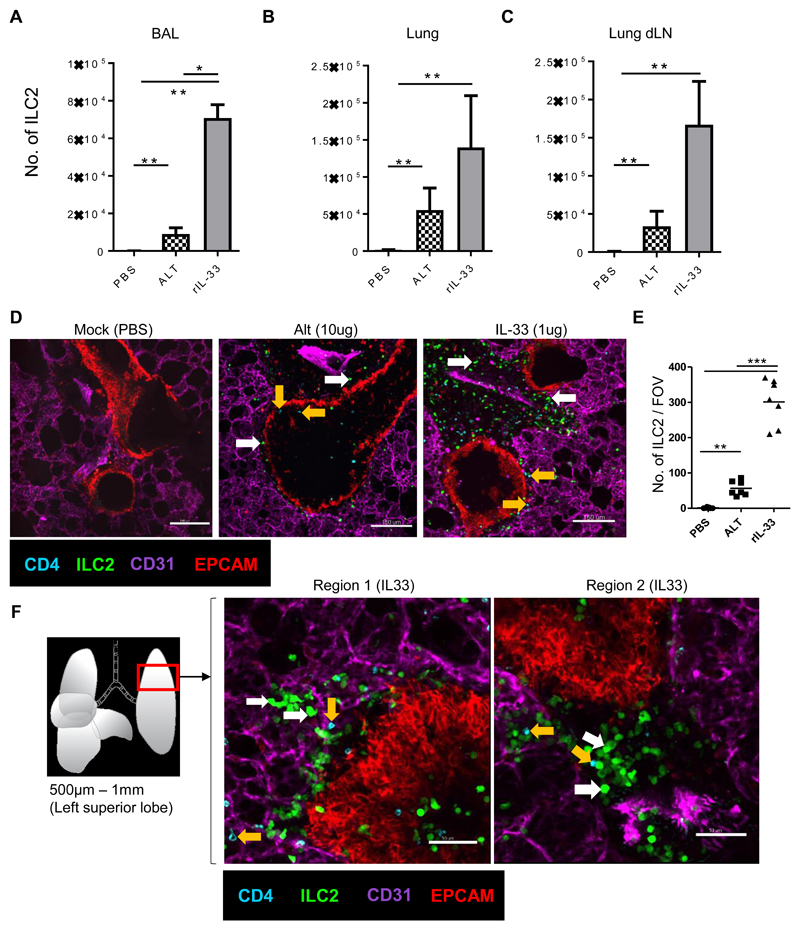
Fig 1. The number of ILC2 rapidly increase in the peribronchial / perivascular region after rIL-33 treatment.
IL13-eGFP mice were treated with 3 doses of rIL-33 (1μg per dose), Alt (10μg) or PBS (25μl) over 1 week and culled 24h after the final dose. The frequency of ILC2 (GFP+ CD45+ Linneg CD3-NKp46-CD127+CD90.2+KLRG1+CD25varIL-13+IL-5+) in the (A) airways (BAL fluid), (B) lung and (C) lung draining lymph nodes (mediastinal). Live viable precision cut lung slices of 200μm thickness were obtained and stained for CD31 (Magenta, the lung structure and blood vessels), CD4 (cyan, T cells, orange arrow), EpCAM (Red, to visualise bronchial epithelium) and GFP (ILC2, white arrow). Images of 1024μm x 1024μm field of view (FOV) were taken under a 20x objective using an inverted confocal microscope. (D) Images showing ILC2 (GFP+CD4-) CD4+ T cells (CD4+GFP-) location in PBS, rIL-33 and Alt treated mice, scale bar, 150 μm. (E) Number of ILC2 (GFP+CD4-) in lung sections per FOV taken under a 10x objective. (F) Schematic illustration of the lung depicting the anatomical location in the lung where precision cut lung slices were prepared. Representative images show two regions of the lung slice from a rIL-33 treated mouse showing distribution of ILC2 and CD4+ T cells, scale bar 150 μm. n = 4 mice per group (Mock(PBS)), n= 6 mice per group (Alt or rIL-33 treatment). Data representative of 4 experiments. *p < 0.05, **p < 0.01, ***p <0.001 and, **** p < 0.0001.

