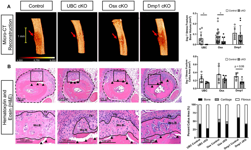
Figure 4. Deletion of VEGFA ubiquitously and in early (Osx+) but not later (Dmp1+) osteolineage cells impaired periosteal bone formation after stress fracture.
A) 3D reconstruction of analysis region shows representative bone formation for each Cre line around stress fracture site (red arrow). UBC cKO stress fractured ulnae showed no quantifiable woven bone via micro-CT. Osx cKO stress fractured ulnae showed impaired woven bone extent and volume. DMP1 cKO stress fractured ulnae showed comparable bone formation to controls. Thresholded bone volume (BV) for entire periosteal callus tissue (which excludes intact cortical bone; Supplemental Figure 3B) on day 7 after stress fracture showed impaired bone formation in UBC cKO and Osx cKO mice versus controls (unpaired t-test; * p<0.05). B) Hematoxylin and Eosin (H&E) transverse sections taken near the crack site (red arrow) of day 7 post stress fractured ulnae displayed a region of woven bone (Wo.B) and expanded fibrous periosteum (Fb; dashed black line) around the stress fracture crack (black arrow heads) for all Cre lines. Quantification of this expanded periosteum area showed that Dmp1 cKO stress fracture calluses trended toward being smaller (p = 0.06) versus controls. UBC and Osx cKO calluses weren’t significantly different in size versus controls. Woven bone (Wo.B), cartilage (Cg) and fibrous (Fb) tissue areas were quantified at higher magnification (Black Box - 20x) and normalized to callus area. No cKO stress fracture calluses had a significantly altered callus composition compared to controls across all Cre lines. Note the minimal amount of cartilage in the stress fracture model.

