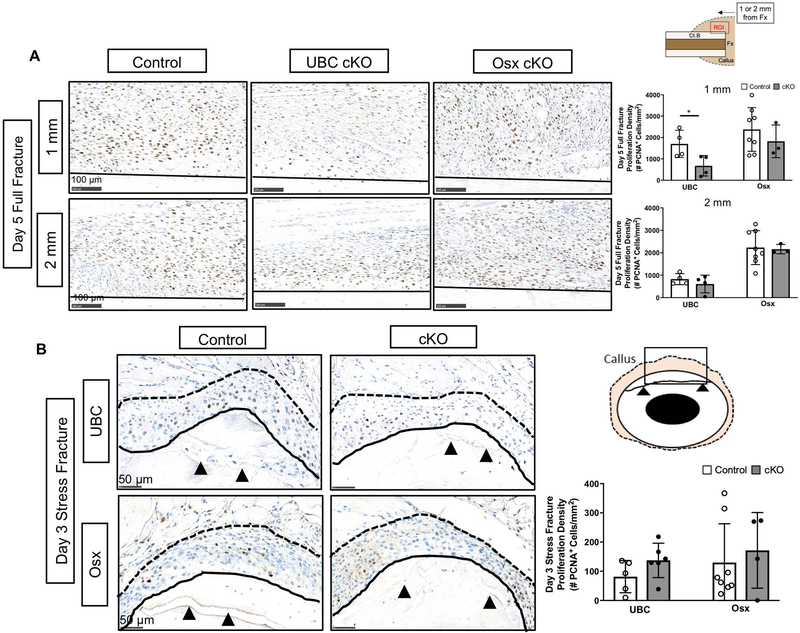
Figure 5. Deletion of VEGFA ubiquitously but not in early (Osx+) osteolineage cells impaired periosteal proliferation after fracture.
A) Representative day 5 full fracture PCNA stained images (20x) of the expanded periosteum at sites 1 mm and 2 mm from the fracture site. The ROI consisted of the region above the cortical bone (black solid line) in each image. PCNA staining of the expanded periosteum 1 mm from the full fracture site revealed a significant reduction in the periosteal proliferative density (# PCNA+ cells/expanded periosteal area) following UBC cKO but not Osx cKO of VEGFA at day 5 (2-Way ANOVA with Sidak Post Hoc Test (* = p < 0.05)). A field of view 2 mm from the full fracture site demonstrated no differences between groups in proliferation density. B) Representative day 3 stress fracture PCNA stained images (40x) showing an enhanced view around the fracture site (black arrows). Stress fracture PCNA staining of the entire expanded periosteum (area between solid black line and dotted black line) after 3 days revealed no reduction in the proliferative density following VEGFA deletion.

