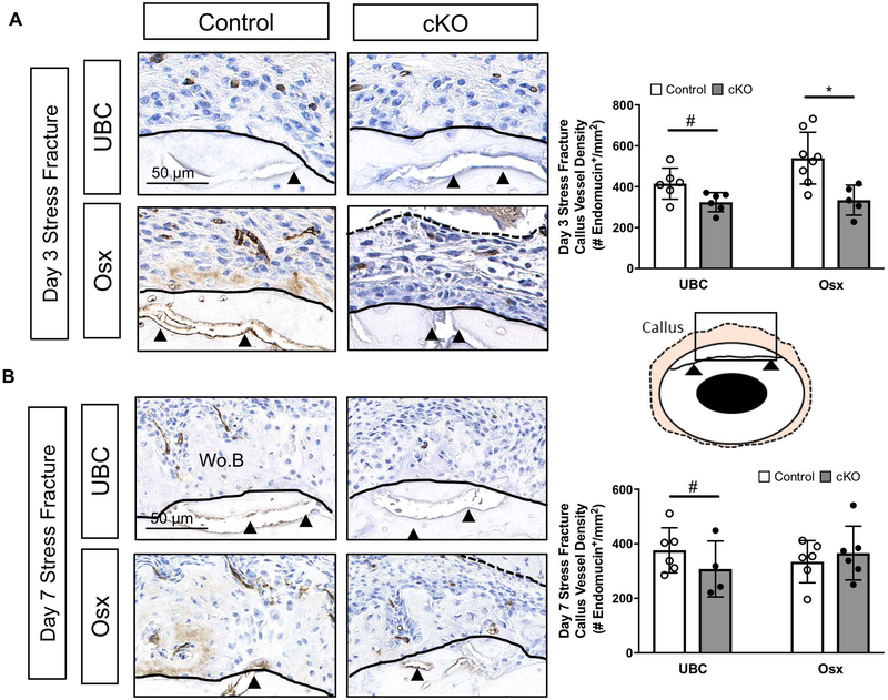
Figure 7. Deletion of VEGFA ubiquitously and in early (Osx+) osteolineage cells impairs periosteal angiogenesis following stress fracture.
A) Representative day 3 endomucin stained images (40x) highlight the region around the stress fracture crack (black arrow heads). The ROI (tan region on schematic) consisted of the expanded periosteal region between the cortical bone (black solid line) and skeletal muscle (dashed black line). Note the absence of appreciable woven bone in all groups at day 3. Endomucin staining quantification of the ROI between genotypes at various timepoints (days 3, 5, 7) revealed a significant decrease in periosteal vessel density (# endomucin+/expanded periosteal area) due to genotype in UBC cKO (2-Way ANOVA, # p<0.05 for genotype) and only at day 3 due to genotype in Osx cKO mice (Sidak Post Hoc Test, * p< 0.05). B) Representative day 7 endomucin stained images (40x) highlight the region around the stress fracture crack (black arrow heads). At day 7, there is now woven bone (Wo.B) infiltrated with blood vessels within the periosteal space. Callus vessel density of the periosteal ROI (# endomucin+/expanded periosteal area) at day 7 (2 way ANOVA; # p-value < 0.05 for genotype).

