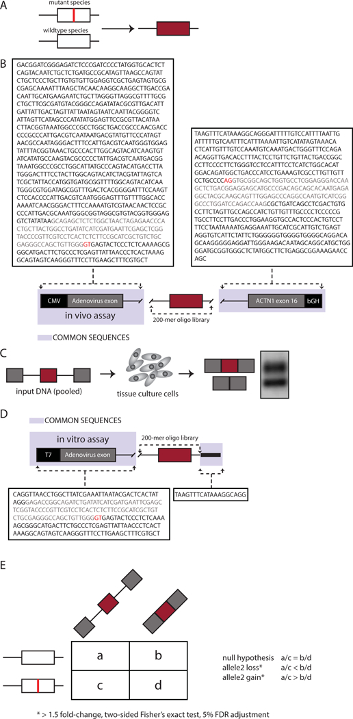
Figure 1: MaPSy challenge.

A. Mutant and wildtype versions of 170-mer genomic fragments flanked by 15-mer common. B. The in vivo splicing reporter consists of the Cytomegalovirus (CMV) promoter and Adenovirus (pHMS81) exon with part of its downstream intron at the 5’ end, followed by the 200-mer oligo library, and exon16 of ACTN1 with part of intron15 and bGH PolyA signal sequence at the 3’ end. C. The in vivo reporters were transfected in hek293 cells. D. The in vitro reporter includes a T7 promoter and Adenovirus (pHMS81) exon. E. Contingency tables were created for each mutant/wildtype pair and include the counts obtained from deep sequencing of the input pool as well as the output-spliced fractions to assess defects in splicing.
