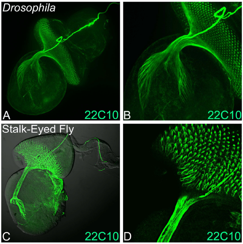
Figure 2. Retinal axon targeting in Stalk-eyed flies and Drosophila.

Monoclonal antibody 22C10 marks all axonal sheath of the photoreceptors in the developing eye. Expression of 22C10 antibody in larval eye antennal imaginal disc of (A, B) Drosophila melanogaster, and (C, D) Stalk-eyed fly. (B) and (D) are magnified images (40×) of A and C. The 22C10 staining marks the axons, axonal targets and its innervation in the brain both in the stalk-eyed fly and Drosophila. Note that all the retinal neurons axons fasciculate into an axonal tract, which innervate the brain of stalk-eyed fly and Drosophila. The orientation of all imaginal discs in the figure is posterior to left and dorsal up.
