Figure 3.
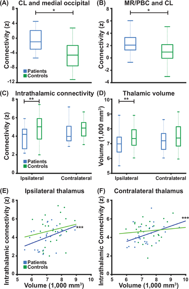
Patients with TLE exhibit perturbed thalamic connectivity and decreased ipsilateral thalamic volume. (A) Patients exhibit loss of negative connectivity between CL intralaminar thalamic nucleus and medial occipital lobe when compared with control subjects. (B) Patients exhibit abnormally increased functional connectivity between CL and MR/PBC as compared with control subjects. (C) Compared with control subjects, patients exhibit decreased intrathalamic connectivity and (D) decreased thalamic volume, on the side ipsilateral to the epileptogenic temporal lobe but not the contralateral side. (E, F) For patients, but not control subjects, higher thalamic volume is correlated with higher intrathalamic connectivity. N=26 patients with TLE before surgery and 26 matched control subjects. *p<0.01 paired t-test, **p<0.05 paired t-test with Bonferroni-Holm correction, and ***p<0.05 Spearman’s Rho with Bonferroni-Holm correction. Centre bar shows median, bottom and top of box designate 25th and 75th percentiles, respectively, and whiskers indicate data extremes. CL: central lateral; MR: median raphe; PBC: parabrachial complex; TLE, temporal lobe epilepsy.

