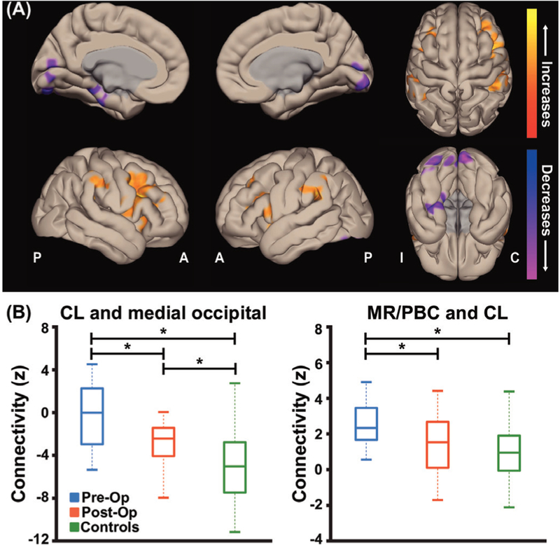
Figure 4.
Postoperative patients with TLE with improved seizures exhibit partial recovery of thalamic arousal network connectivity. (A) Cortical surface views are shown, demonstrating functional connectivity decreases in the medial occipital lobe in postoperative patients with TLE seeded from bilateral thalami. Data represent seed-to-voxel functional connectivity maps (bivariate correlation) comparing fMRI of postoperative versus preoperative patients with TLE (paired t-test, cluster threshold level p<0.05, FDR correction). Two-sided contrasts are shown. fMRIs are orientedwith respect to epileptogenic side for patients with TLE and matched controls were flipped accordingly. (B) Postoperative patients were foundto have some recovery of functional connectivity between CL and medial occipital lobe but connectivity values did not quite reach control subject values (left). However, postoperative patients’ functional connectivity between MR/PBC and CL are decreased compared with their preoperative connectivity with no difference found between postoperative and control subject connectivities (right). N=18 postoperative patients with TLE with improved seizures after surgery, the same 18 patients before surgery, and 18 matched control subjects. *p<0.01 analysis of variance with post-hoc Fisher’s least significant difference procedure. Centre bar shows median, bottom and top of box designate 25th and 75th percentiles, respectively, and whiskers indicate data extremes. A: anterior; C: contralateral; CL: central lateral; FDR: false discovery rate; I: ipsilateral; MR: median raphe; P: posterior; PBC: parabrachial complex; Post-Op: postoperative patients; Pre-Op: preoperative patients; TLE, temporal lobe epilepsy.

