
Figure 1. Amino acid sequence and secondary structure elements of FXN.
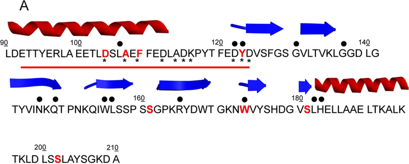
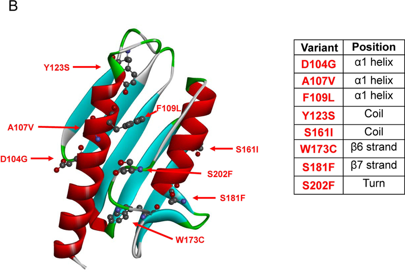
(A) The mutated residues (protein seq. ref. NP_000135.2) object of this study and reported in COSMIC are depicted in bold red, the residues mutated in FRDA are identified by upper black dots. Tyr123 and Trp173 are mutated in FRDA and reported in COSMIC. The lower black stars represent the residues involved in iron binding. The red line highlights the acidic binding region. The secondary structure elements are as reported in 1EKG (Dhe-Paganon et al., 2000). (B) Location of the mutations on FXN structure. Mutated residues are depicted in scaled ball and stick.