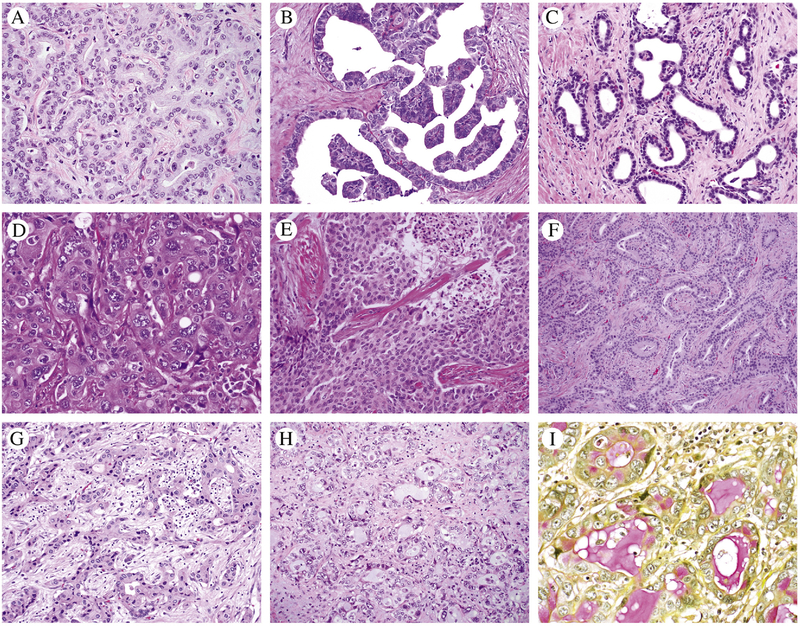
Figure 2.
A-I) Diverse histology and cell shape patterns are seen in IDH1 mutants; A) Anastomosing glands with plump cuboidal cells; B) micropapillary; C) low cuboidal cells with dilated tubules; D) pleomorphic nuclei in polygonal cells, solid pattern; E) Solid pattern; F) Tubular and anastomosing glands, plump cuboidal; G) Anastomosing glnads plump cuboidal; H-I) tubules cuboidal cells and focal with intra and extracellular mucin highlighted by mucicarmine stain.

