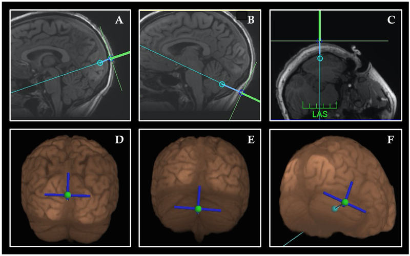
Figure 1: Neuronavigation targets.
(A, D) Sagittal T1-structural image (A) and 3-D reconstruction (D) of visual association area target below the parieto-occipital sulcus. The path of stimulation was angled to pass between the midbrain-pontine junction, angled downward; (B, E) Sagittal T1-structural image (B) and 3-D reconstruction (E) cerebellar vermis target. The path of stimulation was angled to pass between the midbrain-pontine junction, angled upward (C, F) Axial T1-structural image (C) and 3-D reconstruction (F) of lateral cerebellar hemisphere. Twelve subjects received stimulation over the right horizontal fissure shown here; 12 received stimulation over lobule VIII just below (not shown).

