Fig. 1.
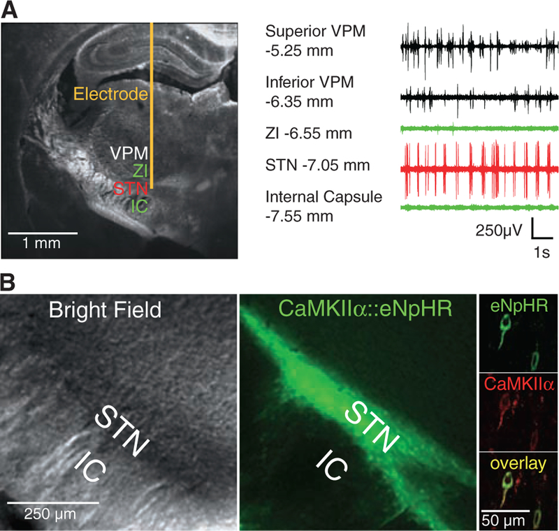
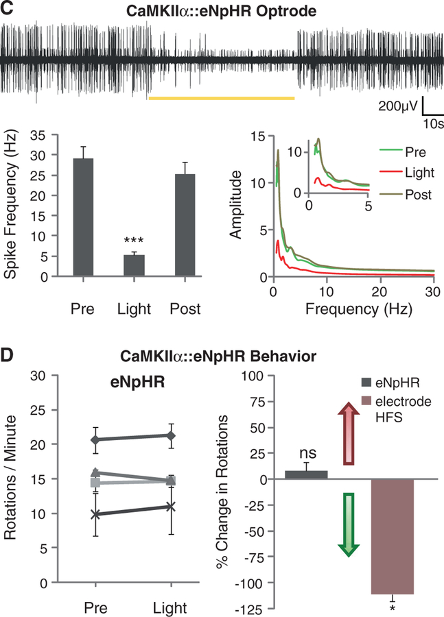
Direct optical inhibition of local STN neurons. (A) Cannula placement, virus injection, and fiber depth were guided by recordings of the STN, which is surrounded by the silent zona incerta (ZI) and internal capsule (IC). (B) Confocal images of STN neurons expressing CaMKIIα::eNpHR-EYFP and labeled for excitatory neuron-specific CaMKIIα (right). (C) Continuous 561-nm illumination of the STN expressing CaMKIIα::eNpHR-EYFP in anesthetized 6-OHDA rats reduced STN activity; representative optrode trace and amplitude spectrum are shown. Mean spiking frequency was reduced from 29 ± 3 to 5 ± 1 Hz (means ± SEM, P < 0.001, Student’s t test, n = eight traces from different STN coordinates in two animals). (D) Amphetamine-induced rotations were not affected by stimulation of the STN in these animals (P > 0.05, n = 4 rats, t test with μ = 0). The red arrow indicates direction of pathologic effects; the green arrow indicates direction of therapeutic effects. The electrical control implanted with a stimulation electrode showed therapeutic effects with HFS (120 to 130 Hz, 60-μs pulse width, 130 to 200 μA, P < 0.05, t test with μ = 0). Percentage change of −100% indicates that the rodent is fully corrected. Data in all figures are means ± SEM ns, P > 0.05; *P < 0.05; **P < 0.01; and ***P < 0.001.


