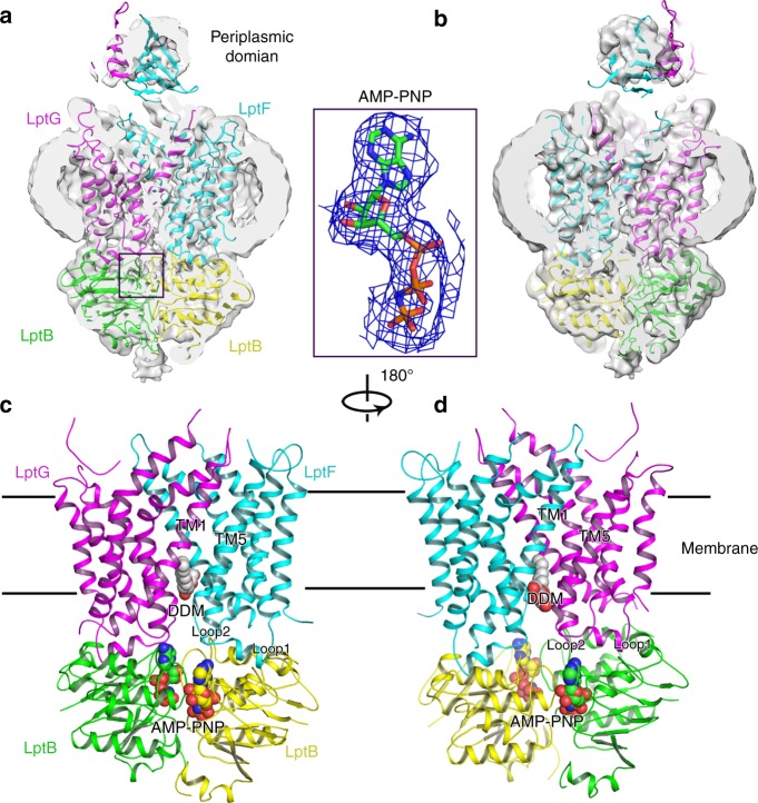
Fig. 4.
Structure of AMP-PNP-bound sfLptB2FGC. a Cryo-EM map of AMP-PNP-bound sfLptB2FGC. The LptC density is not observed. Density of AMP-PNP is shown in blue mesh and AMP-PNP is shown in stick. The colour scheme is the same as Fig. 1. b Rotation of 180° along the y-axis relative to a. c Cartoon representation of AMP-PNP-bound sfLptB2FGC. AMP-PNP is shown in spheres with carbon colour in green or yellow. DDM is shown in sphere with carbon in grey. d Rotation of 180° along y-axis relative to c. The DDM molecule is trapped in the lateral gate TM1F/TM5G

