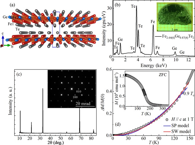
Figure 1.
(a) Crystal structure and (b) X-ray energy-dispersive spectrum of Fe3−xGeTe2 single crystal. Inset shows a photograph of Fe3−xGeTe2 single crystal on a 1 mm grid. (c) X-ray diffraction (XRD) pattern of Fe3−xGeTe2. Inset shows the electron diffraction pattern taken along the [001] zone axis direction. (d) Temperature dependence of the reduced magnetization with out-of-plane field of Fe3−xGeTe2 fitted using spin-wave (SW) model and single-particle (SP) model. Inset shows the temperature dependence of zero-field-cooling (ZFC) magnetization of Fe3−xGeTe2 measured at H = 1 T applied along the c axis.

