Fig. 5.
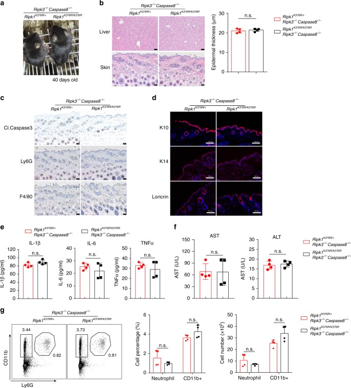
Co-deletion of RIPK3 and Caspase8 fully rescue Ripk1K376R/K376R mice. a Representative macroscopic images of Ripk1K376R/K376RRipk3−/−Caspase8−/− and Ripk1K376R/+Ripk3−/−Caspase8−/− littermate mice at P40. b H&E staining of liver and skin sections of Ripk1K376R/K376RRipk3−/− Caspase8−/− and Ripk1K376R/+Ripk3−/−Caspase8−/− littermate mice at P40 (scale bar, 50 μm), and microscopic quantification of the epidermal thickness from H&E results (Ripk1K376R/+Ripk3−/−Caspase8−/− mice: n = 4; Ripk1K376R/K376RRipk3−/− Caspase8−/− mice: n = 4). c, d Immunohistochemical staining of F4/80, Ly6G, and cleaved Caspase3 (scale bar, 50 μm) (c) or immunofluorescence staining of Loricrin, K10, and K14 (scale bar,100 μm) (d) in skin sections of Ripk1K376R/K376RRipk3−/−Caspase8−/− and Ripk1K376R/+Ripk3−/−Caspase8−/− littermate mice at P40. e Cytokines in liver homogenates were determined with the indicated genotypes at P40 (Ripk1K376R/+Ripk3−/−Caspase8−/− mice: n = 4; Ripk1K376R/K376RRipk3−/− Caspase8−/− mice: n = 4). f AST and ALT in blood were determined with the indicated genotypes at P40 (Ripk1K376R/+Ripk3−/−Caspase8−/− mice: n = 4; Ripk1K376R/K376RRipk3−/− Caspase8−/− mice: n = 4). g Flow cytometry and statistical results of splenocytes stained with Ly6G and CD11b from Ripk1K376R/K376RRipk3−/−Caspase8−/− and Ripk1K376R/+Ripk3−/−Caspase8−/− littermate mice at P40 (Ripk1K376R/+Ripk3−/−Caspase8−/− mice: n = 4; Ripk1K376R/K376RRipk3−/− Caspase8−/− mice: n = 4). CD11b+Ly6G+ cells were identified as neutrophils. In b, e–g, data are mean ± s.e.m. Statistical significance was determined using a two-tailed unpaired t test, n.s., P > 0.05

