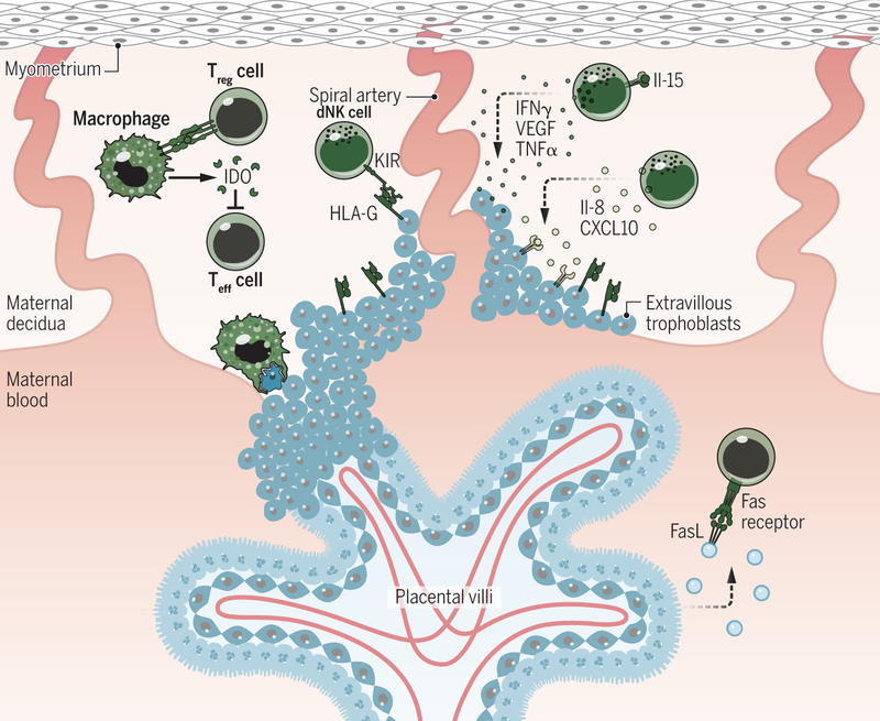
Fig. 3. Mechanisms of maternal tolerance.
The most abundant of the maternal immune cells present in the decidua are dNK cells, which are recruited by various factors released from the decidual stromal cells and placental trophoblasts. The release of uterine IL-15 promotes dNK maturation. The mature dNK cell promotes decidual remodeling and blastocyst implantation through the secretion of cytokines [including IFNγ, VEGF, and tumor necrosis factor α (TNFα)]. The release of IL-8 and CXCL10 by dNK also promotes EVT invasion. dNK cell cytotoxicity is controlled by the binding of HLA-G (expressed on the EVTs) to the inhibitory receptor KIR2DL4. dMØ prevent maternal intolerance by producing IDO, which hinders T cell activation, and phagocytosing apoptotic trophoblasts. Treg cells modulate the activities of both APCs and effector T cells. The syncytiotrophoblast also promotes maternal tolerance by secreting exosomes expressing TRAIL and FasL and by the lack of any MHC expression. Credit: A. Kitterman/Science Immunology

