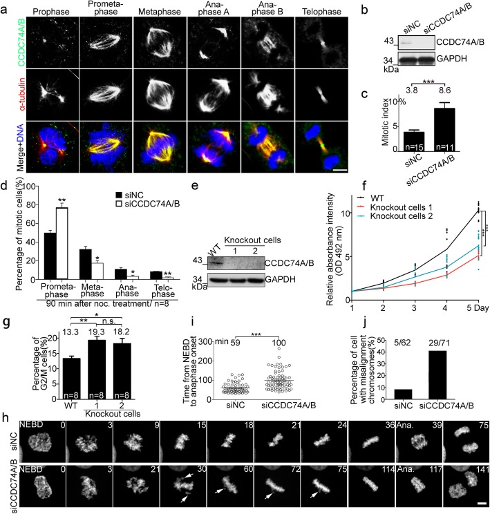
Fig. 1.
CCDC74A/B are localized at mitotic spindles and required for chromosomal alignment. a Immunofluorescence of α-tubulin (red) and CCDC74A/B (green) in COS7 cells. DNA was stained with DAPI (blue). Scale bar, 5 μm. b Western blots of CCDC74A/B in HeLa cells transfected with negative control-siRNA (siNC) or with CCDC74A/B-siRNA (siCCDC74A/B) for 60 h. GAPDH was the loading control. c The mitotic index of HeLa cells after siNC- or CCDC74A/B-siRNA transfection for 60 h (six independent experiments). d Percentages of HeLa cells in mitosis after siNC- or CCDC74A/B-siRNA transfection for 60 h, followed by 1 h nocodazole treatment (noc., 1 μg/ml) then released (6 independent experiments). e Western blots of CCDC74A/B in wild-type (WT) and 2 CCDC74A/B knockout HEK293T cells. GAPDH was the loading control. f Wild-type and 2 CCDC74A/B knockout HEK 293T cells were cultured in 96-well plates. MTT assay was performed at daily intervals over 5 days (6 independent experiments). g Flow cytometric analysis of the percentages of wild-type and 2 CCDC74A/B knockout HEK293T cells in G2/M phase (6 independent experiments). h Time-lapse images of HeLa cells co-transfected with GFP-H2B and either siNC- or CCDC74A/B-siRNA. NEBD, nuclear envelope breakdown; Ana, anaphase. Numbers, time (min) after NEBD. Arrows, misaligned chromosomes. Scale bar, 5 μm. i Time elapsed from NEBD to anaphase onset in the HeLa cells from h (3 independent experiments). j Percentages of mitotic HeLa cells with chromosomal misalignments from h. 5/62, 5 cells with misalignment chromosomes in 62 cells transfected with siNC. 29/71, 29 cells with misalignment chromosomes in 71 cells transfected with siCCDC74B. In c, d, f, and i, data are mean ± SEM (unpaired two-tailed Student’s t test, ***P < 0.001, **P < 0.01, *P < 0.05). In g, data are mean ± SEM (one-way ANOVA test, **P < 0.01, *P < 0.05; n.s., not significant)

