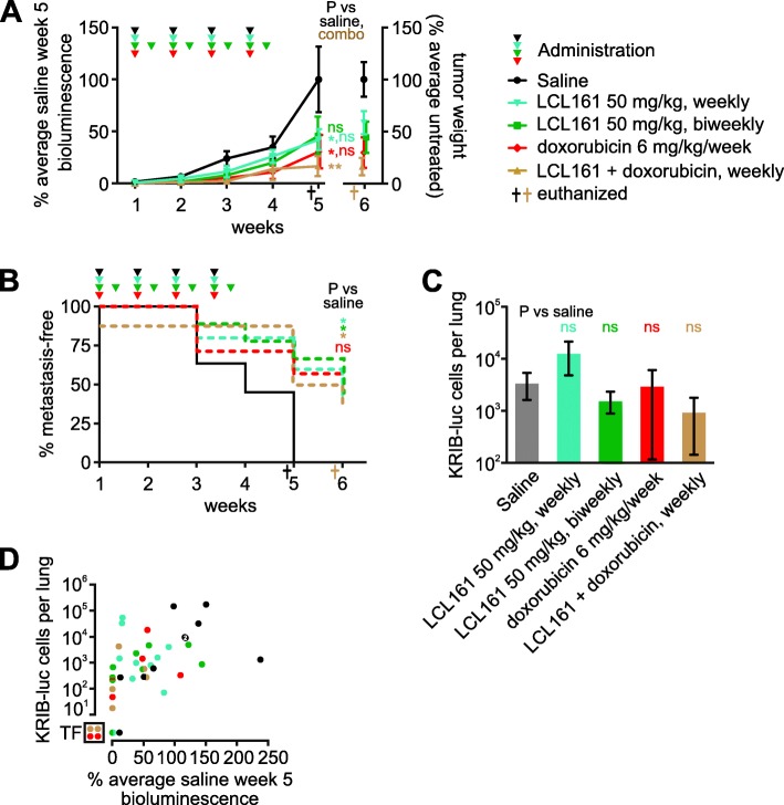
Fig. 6.
LCL161 reduces primary and metastatic growth of human osteosarcoma cells in mice. Luciferase-expressing human KRIB osteosarcoma cells were implanted intramuscularly into nude mice. One to 2 weeks after implantation the mice were administered the specified treatments. a Primary tumor growth was monitored via bioluminescence. Tumor material that could be confidently resected from the surrounding muscle post-mortem was weighed. Some data points at the 5 and 6 week timepoints were slightly horizontally offset to enable all to be visible. A one way ANOVA with Sidak’s post-tests was used to estimate the probability that the drugs significantly affected tumor growth 5 weeks after treatment commenced, and whether the response to co-treatment differed significantly from responses to weekly administration of LCL161 or doxorubicin as sole agents (** P < 0.01; * P < 0.05; ns P > 0.05; n = 7–11, +/− SEM). b The times at which luminescence was first detected in the lungs were recorded. Log-rank (Mantel-Cox) tests were used to compare the onset of metastases in untreated mice versus animals that received each treatment, excluding one co-treated mouse that already had detectable metastatic disease prior to the first treatment. Bonferroni correction was used to adjust the resulting P values for multiple (4) comparisons (* P < 0.05; ns P > 0.05; n = 7–11, +/− SEM). c Lung tumor burden at endpoint was determined by quantitative PCR for surviving mice (excluding one untreated and one-co-treated that had already been euthanized). The assay reliably detected > 10 KRIB-Luc cells per lung. A one way ANOVA with Sidak’s post-tests was used to estimate the probability that the drugs significantly affected lung tumor burden (ns P > 0.05; n = 7–10, +/− SEM). d Metastatic burden was compared with bioluminescence at week 5 (the most reliable measure of primary tumor growth), for each mouse. Data from each mouse is represented by a circle colored to reflect its treatment. Some circles have been cropped to ensure they are all visible. Two saline-treated mice had very similar metastatic and primary tumor burdens; denoted by a white “2” superimposed on those overlapping circles. One saline-treated mouse (denoted by the bottom black dot in the graph) developed detectable lung bioluminescence during the experiment but lacked detectable luciferase DNA within its lungs at the endpoint. Circles in the bottom left box (labeled “TF”) signify tumor free mice. These animals lacked detectable bioluminescence 5 weeks after treatment commenced, no primary tumors were visible upon dissection 1 week later, and they also lacked detectable luciferase DNA in their lungs

