Figure 2:

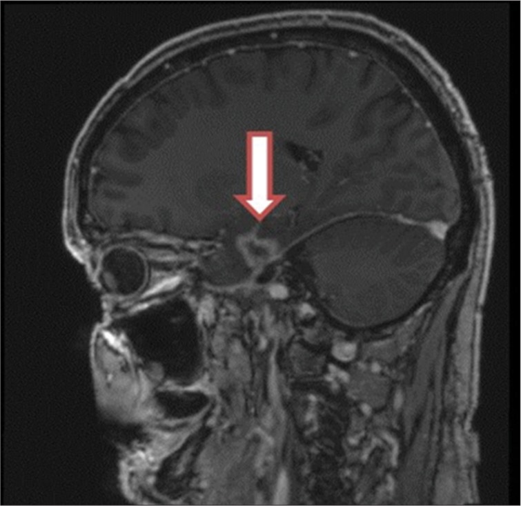
A sagittal view of brain magnetic resonance image with gadolinium enhancement showing temporal lobe necrosis after proton therapy (Courtesy Y. Temel).

A sagittal view of brain magnetic resonance image with gadolinium enhancement showing temporal lobe necrosis after proton therapy (Courtesy Y. Temel).