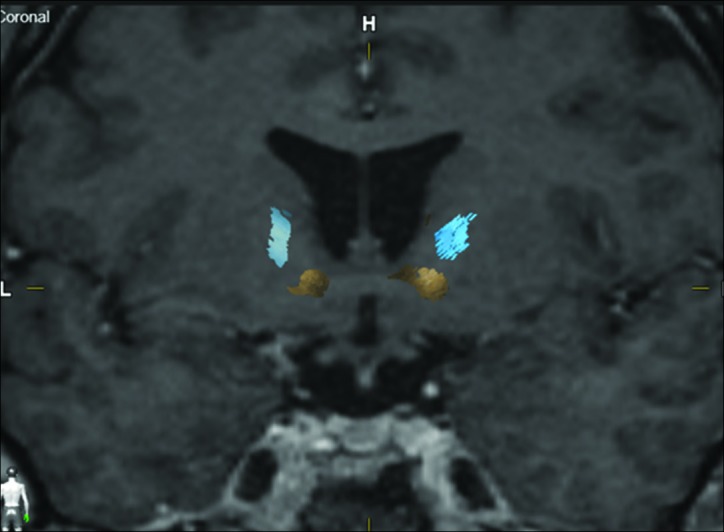
Figure 2:

Coronal magnetic resonance imaging with lateral (blue) and medial (yellow) orbitofrontal cortex (OFC) fiber bundle distribution. Example of a patient with distinct localization of the fibers, lateral OFC bundle cranial to the medial OFC bundle.
