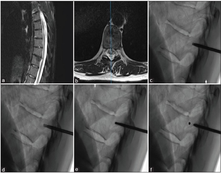
Figure 1:

Fiducial placement procedure. Preoperative sagittal (Panel a) and axial (Panel b) T2-weighted magnetic resonance imaging of the thoracic spine showing a left thoracic disc herniation at T10-T11 in 56-year-old women resulting in spinal cord compression and myelopathy. The patient underwent preoperative pedicle marking under fluoroscopic guidance. Panel c: At the level of the index pedicle, an 11-gauge, 125 mm long trocar is inserted. Panel d: Retrieval of bony fragment with a 13-gauge trepan in the middle portion of the pedicle. Panel e: Two microcoils inserted into the bony defect. Panel f: Reinsertion of the bone cylinder in the defect to lock the fiducial in the pedicle.
