
Figure 4:
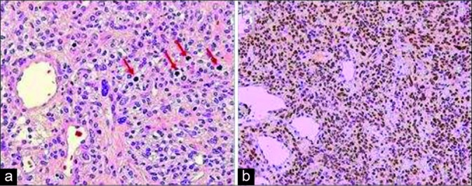
(a) Histologic sections of the left frontal mass demonstrate a high-grade SFT/HPC. Note the four mitotic figures in the mid-upper right (red arrows). (H & E, 200×). (b) Nuclei of the neoplastic cells express STAT6, indicating a fusion of the NAB2 and STAT6 genes (Diaminobenzidine, 100×).