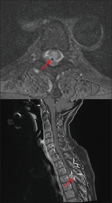
Figure 1:

Upper image: T2 axial image of the lesion surrounding the catheter (red arrow). The granuloma appears as an extra-axial lesion isodense to the myelon. Lower image: T1 contrast sagittal image showing a space-occupying, ring-enhancing, inhomogeneous, extra-axial mass (red arrow) in the spinal canal at the level of T4.
