
Figure 2:
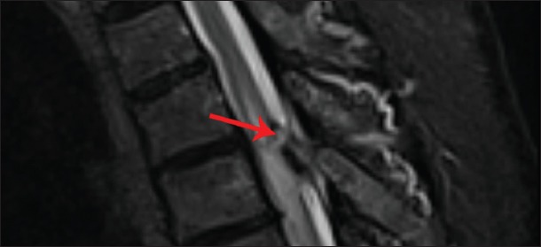
T2 sagittal image of the same lesion with clearly visible extensive T2 signal changes in the spinal cord. The tip of the catheter is seen encased in the granuloma substance (red arrow).
Official websites use .gov
A
.gov website belongs to an official
government organization in the United States.
Secure .gov websites use HTTPS
A lock (
) or https:// means you've safely
connected to the .gov website. Share sensitive
information only on official, secure websites.
T2 sagittal image of the same lesion with clearly visible extensive T2 signal changes in the spinal cord. The tip of the catheter is seen encased in the granuloma substance (red arrow).