
Figure 3:
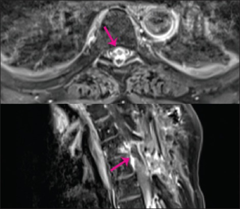
T1 contrast imaging. Upper image: Axial image showing a large recurrence overshadowing and compressing the spinal cord (pink arrow). Lower image: The recurrent granuloma is seen here as a large, ring-enhancing, inhomogeneous, mass-causing high-grade compression of the spinal cord (pink arrow).