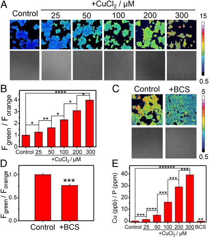
Fig. 2.
Ratiometric fluorescence imaging of labile Cu(I) levels in living cells using FCP-1. (A) Confocal fluorescence microscopy images of HEK 293T cells pretreated with solvent vehicle control or varying concentrations of CuCl2 in complete medium for 18 h. The cells were then washed with PBS, incubated with FCP-1 (5 μM) in DPBS for 45 min, and then imaged. (B) Average cellular ratiometric emission ratios, represented by Fgreen/Forange, as determined from imaging experiments in A performed in triplicate. (C) Confocal fluorescence microscopy images of HEK 293T cells pretreated with solvent vehicle control or BCS (100 μM) in complete medium for 18 h. The cells were then washed with PBS, incubated with FCP-1 (5 μM) in DPBS for 45 min, and then imaged. (D) Average cellular ratiometric emission ratios of FCP-1, Fgreen/Forange, as determined from experiments in C performed in triplicate. A and C are displayed in pseudocolor and referenced to the basal control for that experiment and independently from each other. λex = 458 nm. (E) ICP-MS measurement to determine total cellular 63Cu levels in HEK 293T cells under copper supplementation and depletion relative to the control (with normalization of different cell numbers by total cellular 31P level). Error bars denote standard derivation (SD; n = 3). *P < 0.05, **P < 0.01, ***P < 0.001, ****P < 0.0001, and *****P < 0.00001.

