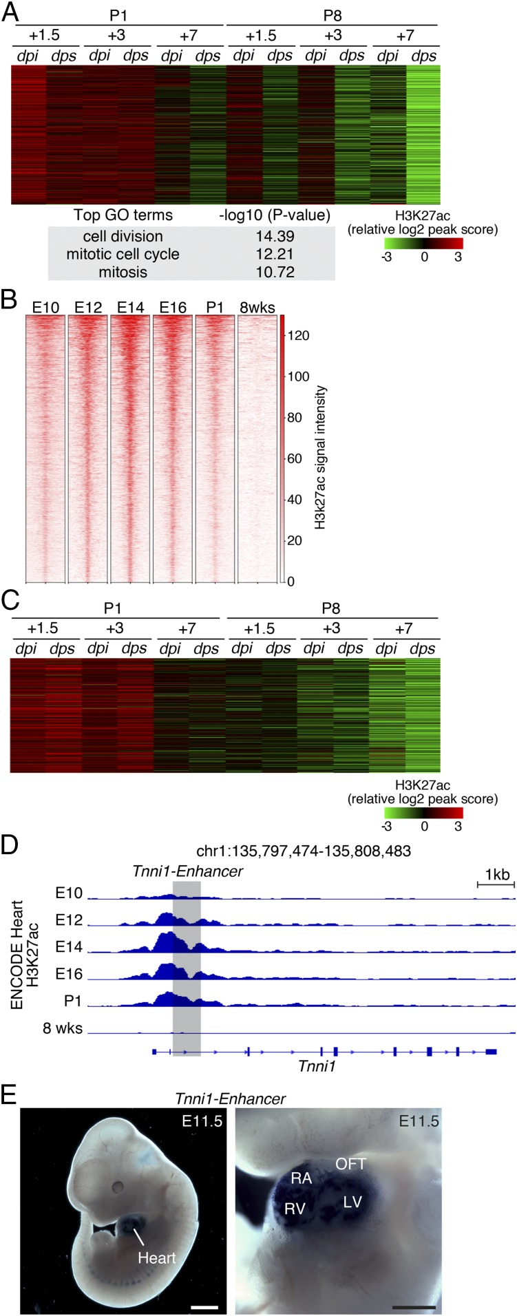
Fig. 4.
Intrinsic developmental gene program is retained in regenerative hearts and is not induced by MI. (A) Heatmap showing the signal intensity of the H3K27ac peaks associated with cell cycle GO terms across all of the samples. (B) Analysis of ENCODE H3K27ac ChIP-Seq data from developing hearts and 8-wk-old hearts identified a developmental gene program composed of active chromatin regions that showed high H3K27ac signals at E10, E12, E14, E16, and P1. (C) H3K27ac signals suggesting the activity of the developmental gene program in hearts subjected to MI or sham surgery at P1 or P8. The developmental gene program activity was retained at the early neonatal stages (P1+1.5 and P1+3) and was not induced by injury. Plotted values are z-scores of normalized peak intensities. (D) ENCODE H3K27ac ChIP-Seq tracks at the Tnni1 gene locus. Tracks representing H3K27ac signals at different embryonic stages are indicated. The region in shadow was cloned and tested for enhancer activity in a transgenic model shown in E. (E) Transgenic enhancer assay showing Tnni1 enhancer-directed heart-specific lacZ expression in E11.5 mouse embryos. (Scale bars: Left, 1 mm; Right, 500 μm.) LV, left ventricle; OFT, outflow tract; RA, right atrium; RV, right ventricle.

