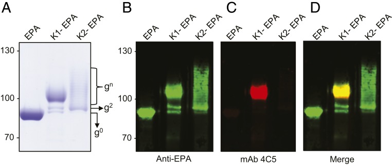
Fig. 3.
K1-EPA and K2-EPA bioconjugate vaccines. (A) Coomassie-stained image of purified EPA, K1-EPA, and K2-EPA. Each lane was loaded with ∼5 µg of glycoconjugate based on total protein. The unglycosylated EPA exists a single band. The K1-EPA exists as multiple glycoforms migrating in smear-like pattern. The K2-EPA bioconjugate migrates with a modal, ladder distribution. Each lane was loaded with ∼0.5 µg of glycoconjugate based on total protein. (B–D) Western blot analysis probing for EPA and the K1 glycan. (B) Anti-EPA Western blot, (C) anti-K1 Western blot, and (D) merge image.

