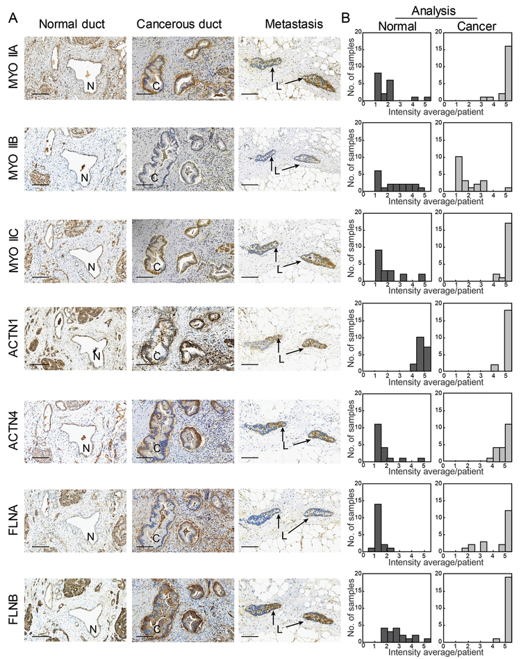
Fig. 2: The mechanoresponsive machinery is elevated in pancreatic ductal adenocarcinoma in human pancreatic tissue.
A. Immunohistochemistry staining of pancreatic tissue from human PDAC patients shows increased expression in the cancerous ducts of mechanoresponsive proteins nonmuscle myosin IIA and IIC, α-actinin 4, and both filamin A and B. Scale bar = 100 μm. For each sample – normal duct (N), cancerous duct (C), and metastatic lesion (L) – the same site is shown across all seven antibodies stained. In addition, both the normal and cancerous ducts are from the same patient. B. Quantification of staining intensity and surface area across 20 patients illustrate an up-regulation of mechanoresponsive proteins, as well as filamin A. Data are plotted as a histogram of the intensity average per patient (described in Fig. S1A and Materials and Methods) across the study group.

