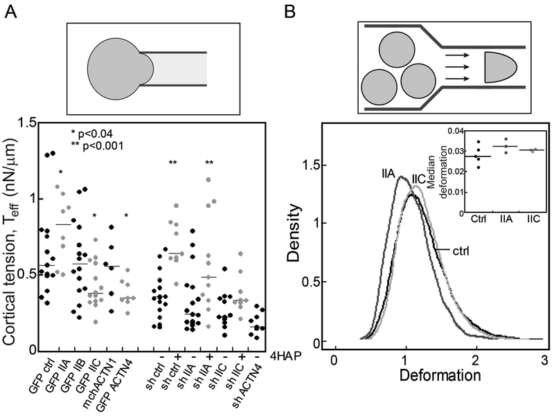
Fig. 4: Mechanoresponsive proteins impact cortical tension and deformability of PDAC cells.
A. The effective cortical tension (Teff) measured by MPA experiments (schematic) is significantly affected by overexpression of myosin IIA, myosin IIC, and α-actinin 4, all mechanoresponsive paralogs. 500 nM 4-HAP increases the cortical tension of sh-control cells. 4-HAP also increases the cortical tension in myosin IIA knockdown cells, whose primary myosin paralog myosin IIC is activated by 4-HAP (42). Knockdown of myosin IIC also leads to a decrease in cortical tension, unchanged by 4-HAP challenge. Medians plotted; * p<0.03; **p<0.0001 relative to control. B. RT-DC experiments (schematic) demonstrate increased deformation when myosin IIA and myosin IIC are knocked-down (plotted as a probability distribution). All cell lines are generated from Panc10.05 cells. N=7521 cells for control, 9632 cells for myosin IIA knockdown, and 5933 cells for myosin IIC knockdown. Cell types are distinct (p<0.0001). Inset: Median deformation of three to five RT-DC independent runs across cell types from which the elastic modulus was calculated. Average median deformation values are 0.027±0.002 (mean±SD) for ctrl, 0.032±0.002 for IIA knockdown, and 0.031±0.0003 for IIC knockdown.

