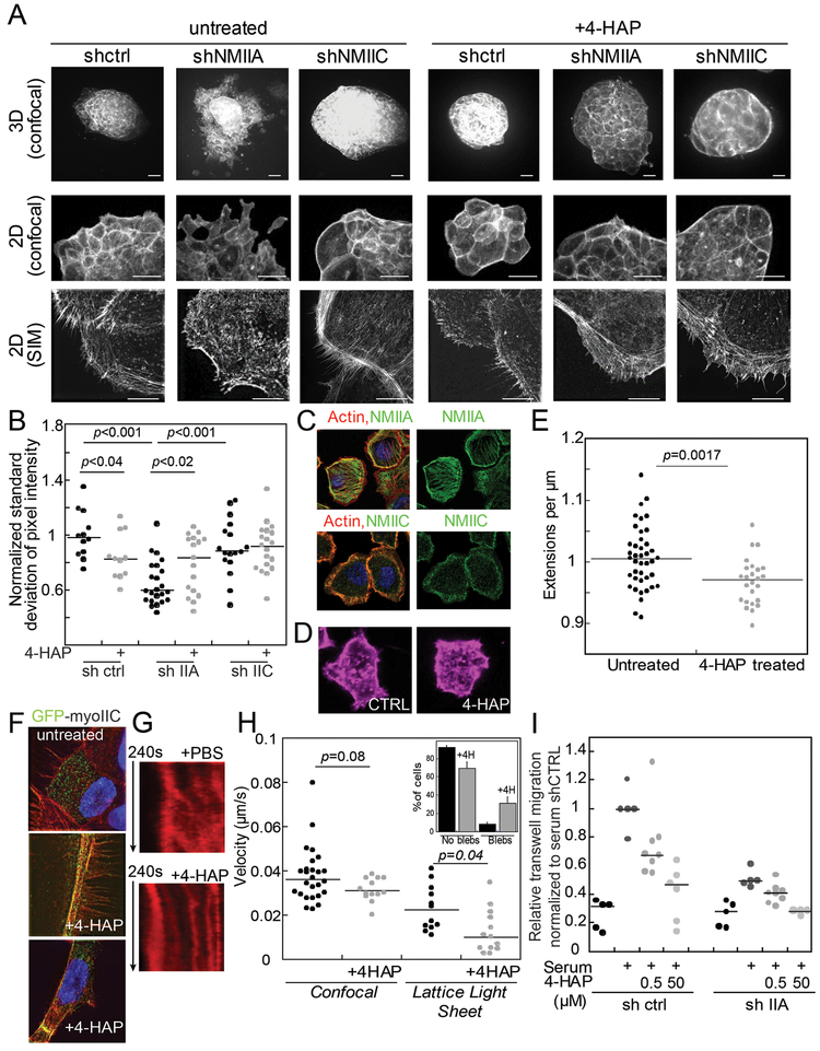
Fig. 5: Myosin IIC alters cytoskeletal actin architecture and cell behavior upon 4-HAP treatment.
A. Panc10.05 tissue spheroids grown, stained with phalloidin, and imaged in 3D (Matrigel) or 2D (collagen) show dissemination, exacerbated in myosin IIA knockdown. Myosin IIC depletion suppresses dissemination and generates actin cortical belts. 4-HAP induces actin belts in control and myosin IIA-depleted spheroids. These belts are already present in myosin IIC-depleted spheroids. Scale bar, 40 μm (confocal); 10 μm (SIM). B. Quantification of these actin structures using normalized standard deviation of pixel intensity; see Methods. Medians plotted; p values on graphs. C. Endogenous myosin IIA in AsPC-1 cells localizes on actin stress fibers; myosin IIC localizes to the actin cortex. D. 4-HAP reduces the number of extensions formed by AsPC-1 cells. E. Numbers of extensions/μm of perimetric distance are plotted. F. GFP-myosin IIC decorates actin filaments, especially actin belts induced by 4-HAP in tissue spheroids. G. Sample kymographs of line scans across active leading edges in AsPC-1 SirAct live-stained cells. H. 4-HAP decreases retrograde flow. Medians are plotted. Inset shows increase in bleb formation in 4-HAP-treated cells (p=0.0028). I. AsPC-1 shCTRL and shIIA cells show dose-dependent reduction of transwell migration upon 4-HAP treatment. Medians are shown.

