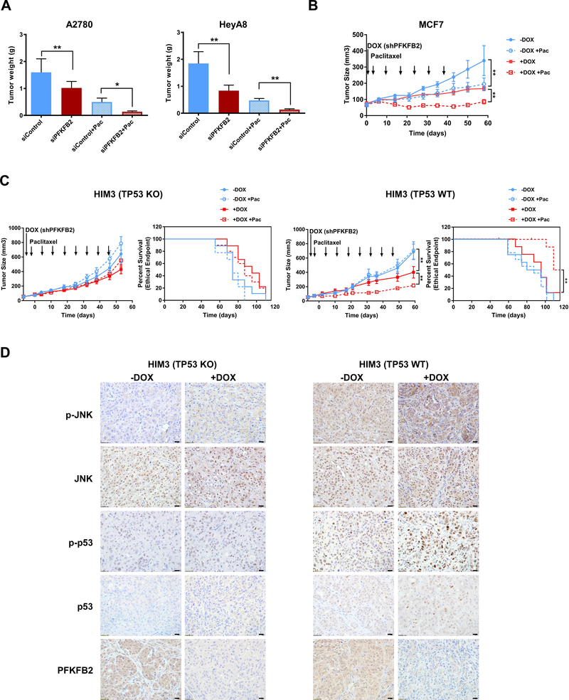
Figure 6. Knockdown of PFKFB2 significantly increases paclitaxel sensitivity in ovarian and breast cancer xenografts.
(A) Tumor growth of ovarian cancer cells in female athymic nude mice after treatment with PFKFB2 siRNA-DOPC with or without paclitaxel. 1 million HeyA8 or A2780 cells were injected intraperitoneally (ip). After 7-day inoculation, mice (n=10) were treated with siRNA-DOPC at 5 ug per mouse twice per week and/or paclitaxel at 30 ug per mouse once a week for 4 weeks. All mice were sacrificed and tumors were weighed when mice in control group became moribund. Tumor growth by weight under different treatments was plotted as mean ± S.D. (*p<0.05; ** p<0.01). (B) Tumor growth of MCF-7 breast cancer cells in female athymic nude mice. 3 × 10⁶ cells were injected into the fourth mammary fat pads three days after estradiol pellet implantation. Tumor-bearing mice were randomized into 4 treatment groups (n=7) when tumors reached 50 mm3. Mice were treated with 1. Sucrose water (-DOX); 2. Sucrose water and 5 mg/kg paclitaxel (-DOX + Pac); 3. Doxycycline water (+DOX) and 4. Doxycycline water and 5 mg/kg paclitaxel (+DOX + Pac). All mice were sacrificed after 6 weeks. (** p<0.01). (C) Tumor growth of HIM3 isogenic breast cancer cells in NOD/SCID mice. 1X10⁶ tumor cells was injected into the fourth mammary fat pads of mice. After tumors reached 50 mm3, tumor-bearing mice were randomized into 4 groups (n=8). Mice were treated as described in Fig. 7B for 8 weeks. Animal survival was evaluated from the start of treatment until tumors reached 1000 mm3. Survival curves were generated by GraphPad Prism 7. (*p<0.05; ** p<0.01). (D) Representative images of IHC with indicated antibodies from HIM3 TP53 KO tumor tissue and HIM3 TP53 WT tumor tissue. –DOX: wild-type PFKPB2, +DOX: shPFKFB2, scale bar: 20μM.

