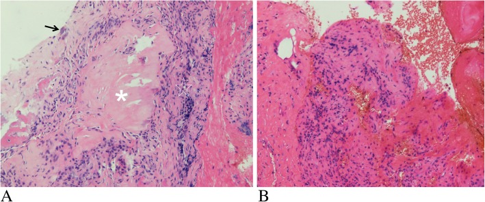
Fig. 4.
Histological section of a surgically resected specimen of the same patient. Hematoxylin and erosin (HE) staining (a and b) showing the massive deposition of uric acid crystals (a, asterisk) surrounded by granulomatous inflammation (a, pentagon), and few small calcium deposition (a, arrow), as well as fiber and angiogenesis with a large number of chronic inflammatory cells infiltration (b)

