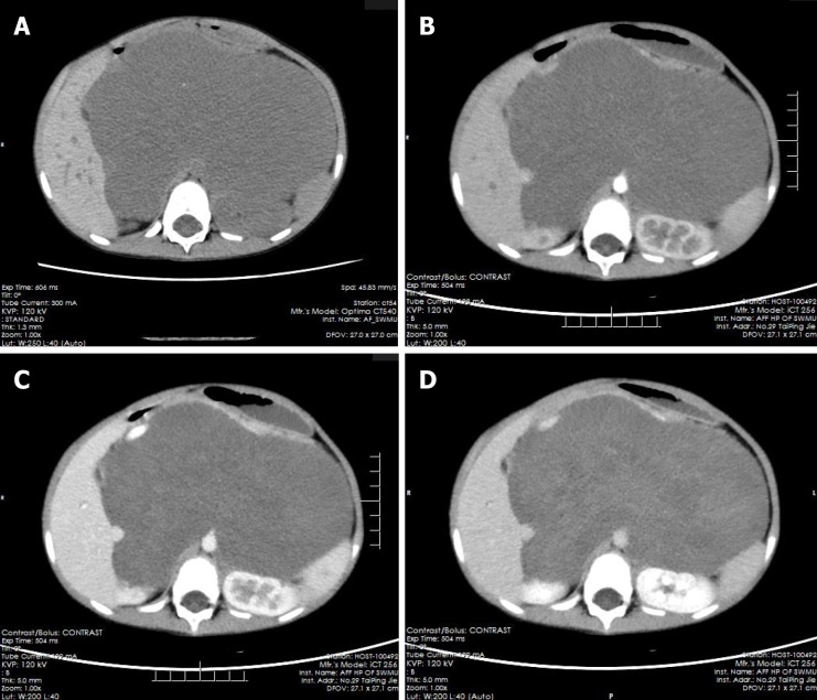
Figure 1.
Multiphase enhanced computerized tomographic images of the patient. A: Giant retroperitoneal hypodense mass in pre-contrast and punctate calcification could be noted; B: The hypodense mass without enhancement in arterial and venous phase, and the stomach and kidneys were compressed and displaced; C: The mass presented mild enhancement in venous phase; D: The mass showed patchy and flocculent enhancement in delay phase, and the extent of enhancement was greater than that of the venous phase.

