Figure 1.

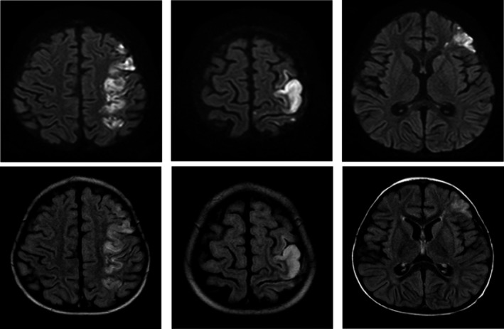
Magnetic resonance imaging of abnormal restricted diffusion/infarct within the left middle cerebral artery and watershed distribution (day 4 of hospitalization). DWI (top) and T2 FLAIR (bottom)

Magnetic resonance imaging of abnormal restricted diffusion/infarct within the left middle cerebral artery and watershed distribution (day 4 of hospitalization). DWI (top) and T2 FLAIR (bottom)