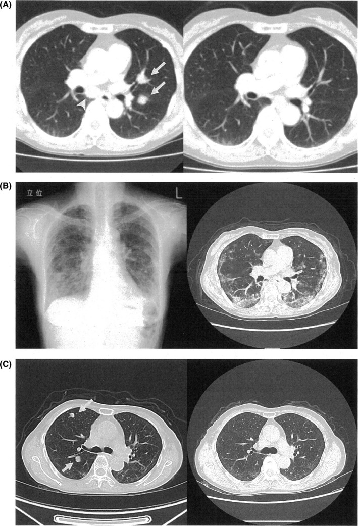
Figure 2.

Chest images of the patient. A, Axial images of the contrast‐enhanced computed tomography (CT) at the main bronchus level. Left panel: image before nivolumab therapy; metastatic tumors visible in mediastinum [arrow‐head] and left lung [arrow]; Right panel: images after injecting nivolumab three times; metastatic tumors have disappeared. B, Left panel, chest X‐ray; Right panel, contrast‐enhanced CT image at main bronchus level after injecting nivolumab 24 times in April 2018; Reticulo‐nodular shadows visible in lower lung fields. C, axial images of the contrast‐enhanced CT at the subcarina level. Left panel: image in August 2018 shows reappearance of multiple lung metastases (arrow); Right panel: image in November represents disappearance of metastatic tumors
